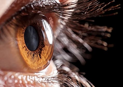
 The eye is one of the most complex structures in the human body, second only to the brain. The eye has over two million moving parts.
It carries the visual data crucial for our survival and education. More than half of the brain’s neocortex is completely devoted to processing this visual data.
Most of us are familiar with the external anatomy of the eye. These include the eyelids, lashes, iris, the whites of the eye, and the pupil.
We may even know that the pupil plays a role like a camera shutter, adjusting to different lighting. You may be familiar with the conjunctiva.
This is the protective mucous membrane near our tear ducts. It becomes inflamed and itchy during allergy season! But the external anatomy goes far beyond what we can see.
What is the cornea?
One of the key players in the eye’s external anatomy for vision problems and correction is the cornea. The cornea is the dome-shaped piece that covers the iris and the pupil.
Many people mistake the cornea for the lens of the eye, but they’re not the same. Structurally, the cornea is on the outside of the pupil and the iris, while the lens is underneath them.
The cornea and lens work together to refract data onto the retina. But it’s the cornea that does two-thirds of all the refractive heavy-lifting!
A few of the common corneal diseases that we treat at Huffman & Huffman, P.S.C. include keratoconus, Fuchs’ Dystrophy, and corneal infections.
